Hexokinase catalyzes the first step of glycolysis (the synthesis of glucose-6-phosphate from glucose and ATP.
Hexokinase is a monomeric enzyme consisting of two conserved domains, each an actin like ATPase domain. Isoforms of this enzyme from different species retain this conserved structure despite having as little as 30% amino acid identity between them.
Binding of glucose causes the two halves of the enzyme to close around the active site like a clam shell. This shields the transfer of the phosphate group from exposure to water, which could result in simple hydrolysis of the ATP instead of phosphoryl group transfer to glucose.
Phosphoglucose isomerase catalyzes the isomerization of glucose-6-phosphate to fructose-6-phosphate.
The reaction proceeds by catalyzing the opening of the glucose ring (mediated by His 388) and the transfer of a proton from C2 to C1 through an enediol intermediate. Lys 518 and Thr 214 are involved in the proton transfer reaction.
Phosphoglucose isomerase is a homodimer with the two subunits embracing each other. The protein is predominantly α helical with some short stretches of β sheet and is classified as an α/β protein. The only domain found in this protein is a SIS domain, an αβα sandwich with the β sheet containing 5 parallel strands.
Phosphofructokinase catalyzes the addition of the second phosphate to the carbohydrate substrate (fructose-6-phosphate) in glycolysis. This reaction requires ATP and unlike many of the reactions of glycolysis, it is not reversible under physiological conditions. Phosphofructokinase is also one of the most highly regulated steps of glycolysis.
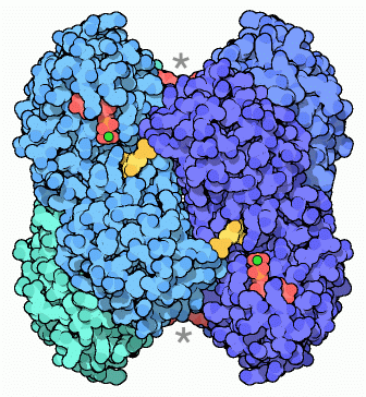
In microbes phosphofructokinase is a homotetramer. Each of the four subunits has a catalytic site (shown by a star in the image below) as well as a different regulatory site where allosteric regulators bind (shown in red and gold).
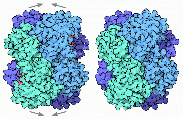
The entire complex alters conformation upon binding of allosteric regulators, fluctuating between an open catalytically active R state and a condensed, inactive T state.
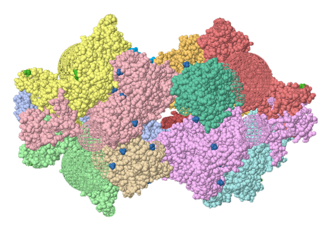
The structure of eukaryotic phosphofructokinase is much more complicated than the bacterial enzyme. The holoenzyme from yeast (Pichia pistoris) is a dodecamer with A4B4C4 stoicheometry weighing in at nearly a million daltons.
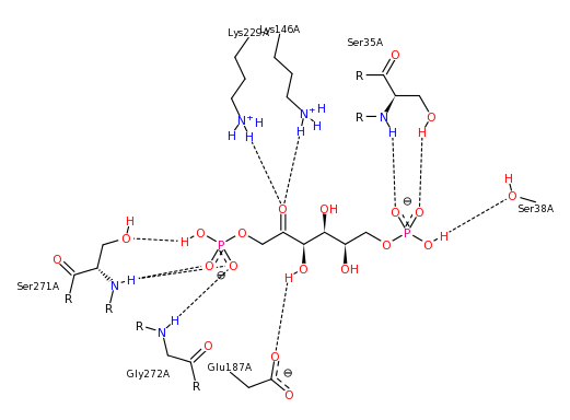
Aldolase catalyzes the cleavage of a hexose phosphate (fructose-1,6-bisPhosphate) into two triose phosphates (dihydroxyacetone phosphate and glyceraldehyde-3-phosphate). Binding of substrate occurs through numerous polar interactions (hydrogen bonds and salt bridges) with the substrate binding site.
The mechanism of the reaction employs a Schiff base formed between the substrate and a lysine side chain (Lys 229). An aspartate residue (Asp 33) aids in the cleavage step by deprotonating the hydroxyl on carbon 4.
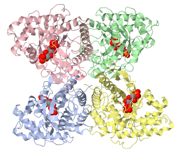
Structurally, aldolase is a mixed α/β structure consisting of a single beta sheet that cups the substrate and helps form the active sheet. This fold is termed a TIM barrel and is observed in several other proteins that bind carbohydrates. 11 α-helices and 3 short 3/10 helices complete the monomeric structure.
The structure found in the cell (the biological assembly) is a homotetramer consisting of 4 of these monomers in a tetrahedral arrangement with the active sites oriented towards the middle of the structure.
Lactate dehydrogenase reduces pyruvate to lactate and in the process oxidizes NADH back to NAD+ conditions. In Eukaryotes, lactate dehydrogenase is a tetrameric enzyme. Animals have two separate genes that code for two separate polypeptides that generate either the muscle (M) or heart (H) isoforms.
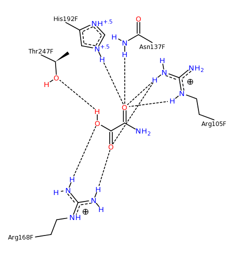
Different combinations of M and H isoforms (e.g. H3M1) lead to different isozymes and different activities in different tissues. This difference in activity is due to a difference in Km for pyruvate which is dictated by the active site histidine (His 192). The pKa of this histidine differs by as much as 0.94 pH units between the muscle and heart isoforms, and results in the different kinetic s observed
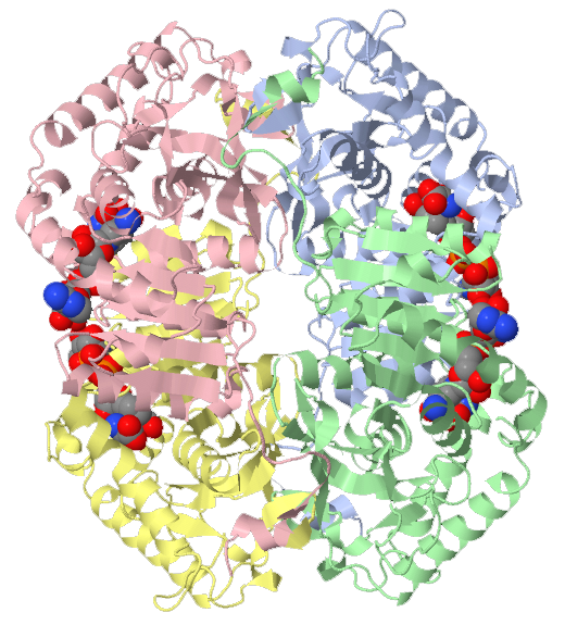
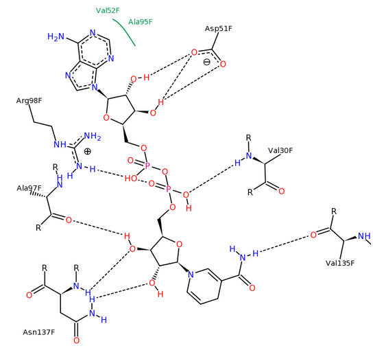
Lactate dehydrogenase is a mixed α/β protein and consists of a mixture of helix and sheet. Each subunit of lactate dehydrogenase folds into two discrete domains. The amino terminus forms what is known as a Rossman fold, a conserved domain found in many proteins that bind to NAD+ or NADP+. The carboxy terminus folds into a domain known as a lactate dehydrogenase fold, a domain conserved among all lactate dehydrogenases that binds to pyruvate.
Over 140 structures of lactate dehydrogenase have been determined. Many of these structures are bound to inhibitors of the enzyme. Plasmodium falciparum, the parasite that causes malaria uses lactate dehydrogenase to survive in the erythrocyte. This enzyme has been identified as a target for possible drug treatments for malaria, hence the interest in the structure of the complexes.
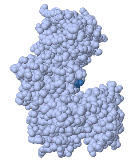
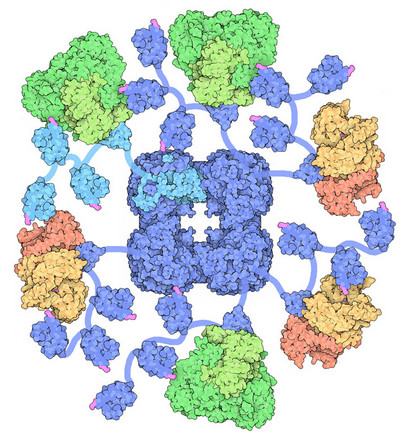
Pyruvate dehydrogenase is a large, multi-subunit complex which catalyzes the conversion of pyruvate to acetyl-CoA. The complex catalyzes three different reactions and uses three different subunits to catalyze the reaction. The reactions are linked by the means of a lengthy and flexible dihydrolipoyllysine arm. This arm can reach between the different subunits to shuttle reactants from one active site to the next.